- These photos contain views of the Morgellon's disease structure know
as the Callus.
-
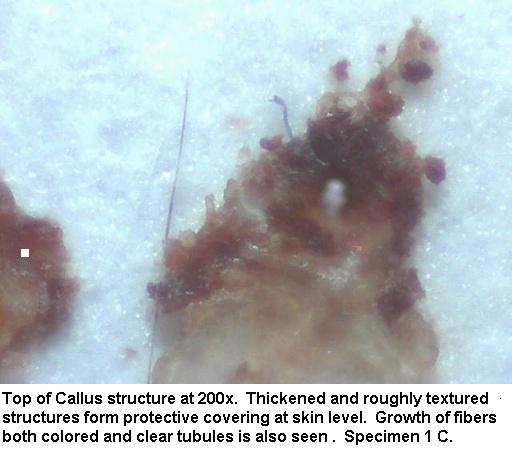
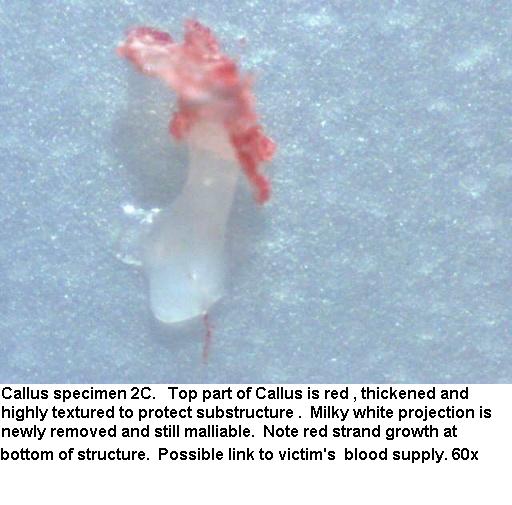
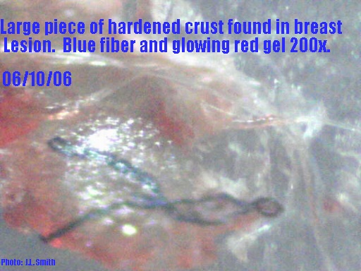
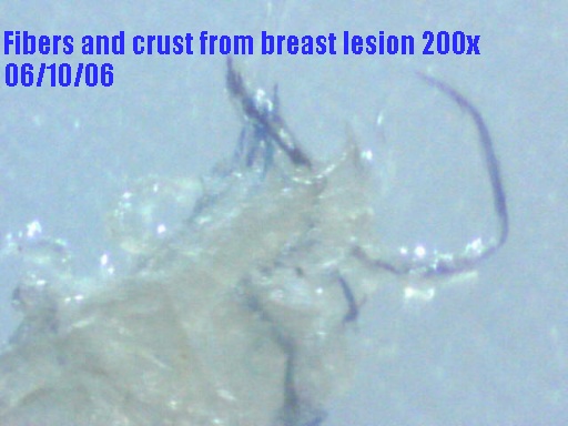
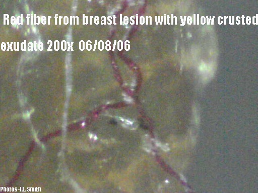
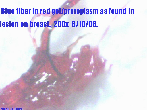
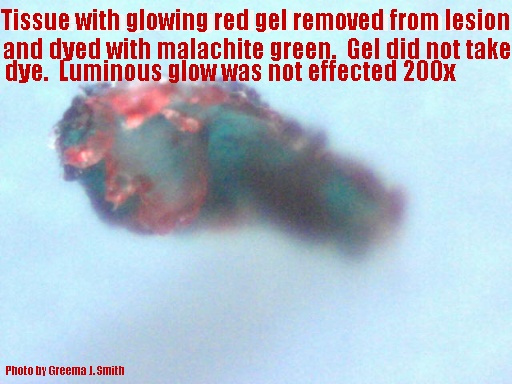
- There are photos which show the nearly impermeable thickened,
reddish-callused component that is visible from the surface of the
skin. This callus protects the substructures and continues to
grow as well. The photos also show other structures associated
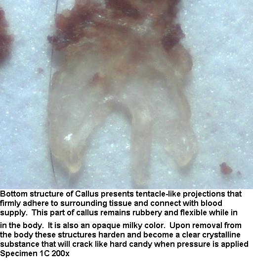
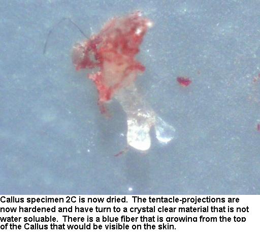
with that part of the Callus. There are other photos which show the milky
to transparent Callus-tentacles and the appurtenant structures associated
with that subcutaneous portion of the Callus.
-
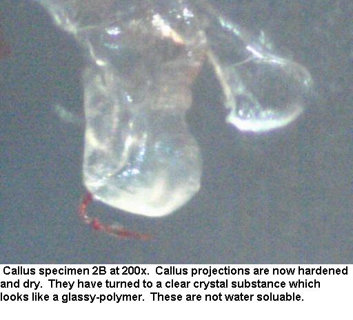
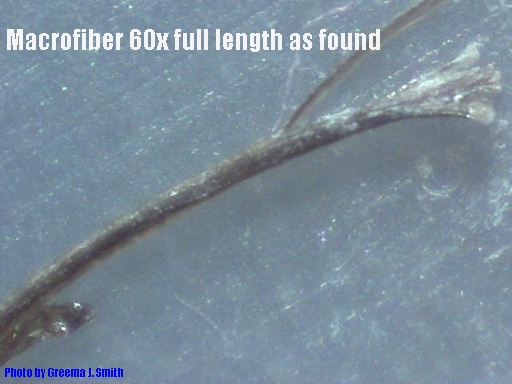
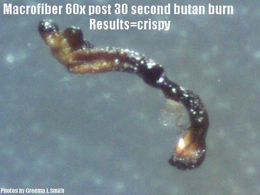
- There is a marked difference in the clarity and texture of the callus
materials before and after removing them from the body. I have shown
these differences as best I can.
-
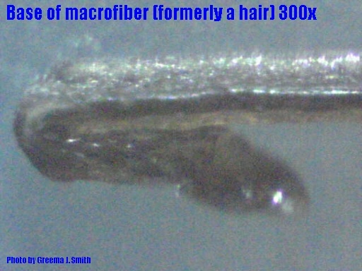
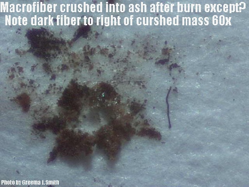
- It is important to not that many of these structures are tightly
grouped together forming a nearly intractable fortress to protect the feeding
Callus structures beneath the skin. If even a single one
is these structures is able to be removed from the Callus it is with
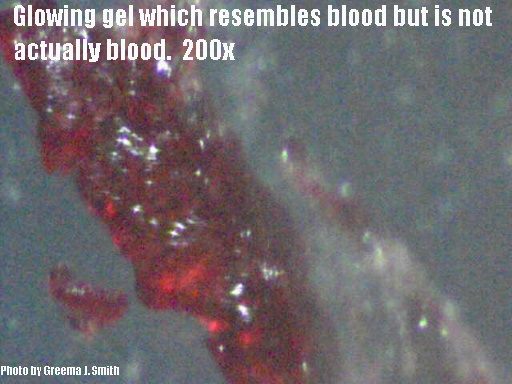
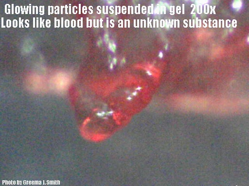
great pain and great physical force. In the aftermath there is profuse
bleeding of an unusually thinned watery version of normal blood.
It is as if some chemical compound in the tentacle is acting as a
blood thinner. The removal of these callus particles leaves
a deep hole in the skin which will generate and fill the void with
a new tentacled protrusion is just hours.
|